An important new study indicates that calcium deposits in the eye cause Age-related Macular Degeneration. Little by little the deposits build up until drusen, hard deposits of fat and protein, can be detected. Until this study, no one knew how drusen was formed. AMD is characterized by lots of drusen, which show up in the retinal scans recommended in BELLA, the LivingTheCRWay Alzheimer’s/Brain/Aging gauge project.
Here’s a quote from one of the researchers:
“The calcium-based spheres (deposits) are made up of the same compound that gives teeth and bone their strength, so removal may not be an option,” says Dr. Lengyel. “However, if we could get to the spheres before the fat and protein build-up, we could prevent further growth.”
A goal of the CR Way is to minimize or prevent or deposit formation. Maintaining optimal vitamin D status, reasonable dietary calcium intake, regular nitric oxide influx, and limiting dietary oxalates may all reduce formation of these calcium deposits.
Note that some of the calcium deposits( spheres) were coated with beta-amyloid, the characteristic protein found in excess in Alzheimer’s
Here is the press release from the University College London, where the study was conducted:
Major cause of blindness linked to calcium deposits in the eye
19 January 2015 University College London – UCL
Microscopic spheres of calcium phosphate have been linked to the development of age-related macular degeneration (AMD), a major cause of blindness, by UCL-led research.
AMD affects 1 in 5 people over 75,causing their vision to slowly deteriorate, but the cause of the most common form of the disease remains a mystery.* The ability to spot the disease early and reliably halt its progression would improve the lives of millions, but this is simply not possible with current knowledge and techniques.
The latest research, published in Proceedings of the National Academy of Sciences, has implicated tiny spheres of mineralized calcium phosphate, ‘hydroxylapatite’, in AMDprogression. This not only offers a possible explanation for how AMD develops, but also opens up new ways to diagnose and treat the disease.
AMD is characterized by a build-up of mainly protein and fat containing deposits called ‘drusen’ in the retina,which can prevent essential nutrients from reaching the eye’s light-sensitive cells,‘photoreceptors’. Photoreceptors are regularly recycled by cellular processes,creating waste products, but drusen can trap this ‘junk’ inside the retina,worsening the build-up. Until now,nobody understood how drusen formed and grew to clinically relevant size.
The new study shows that tiny calcium-based hydroxyapatite, commonly found in bones and teeth, could explain the origin of drusen. The researchers believe that these spheres attract proteins and fats to their surface, which build up over years to form drusen. Through post-mortem examination of 30 eyes from donors between 43 and 96 years old, the researchers used fluorescent dyes to identify the tiny spheres, just a few microns – thousandths of a millimeter – across.
“We found these miniscule hollow spheres inside all of the eyes and all the deposits that we examined, from donors with and without AMD,”explains Dr Imre Lengyel, Senior Research Fellow at the UCL Institute of Ophthalmology and Honorary Research Fellow at Moorfields Eye Hospital,who led the study. “Eyes with more of these spheres contained more drusen. The spheres appear long before drusen become visible on clinical examination.
“The fluorescent labeling technique that we used can identify the early signs of drusen build-up long before they become visible with current methods. The dyes that we used should be compatible with existing diagnostic machines. If we could develop a safe way of getting these dyes into the eye, we could advance AMD diagnoses by a decade or more and could follow early progression more precisely.”
Some of the mineral spheres identified in the eye samples were coated with amyloid beta, which is linked to Alzheimer’s disease. If a technique were developed to identify these spheres for AMD diagnosis, it may also aid early diagnosis of Alzheimer’s. Whether these spheres are a cause or symptom of AMD is still unclear, but their diagnostic value is significant either way. As drusen are hallmarks of AMD, then strategies to prevent build-up could potentially stop AMD from developing altogether.
“The calcium-based spheres are made up of the same compound that gives teeth and bone their strength, so removal may not be an option,” says Dr Lengyel.“However, if we could get to the spheres before the fat and protein build-up,we could prevent further growth. This can already be done in the lab, but much more work is needed before this could be translated into patients.”
“Our discovery opens up an exciting new avenue of scientific research into potential new diagnostics and treatments, but this is only the beginning of a long road.” says Dr Richard Thompson, the main international collaborator from the University of Maryland School of Medicine, USA.
Here is the study:
Identification of hydroxyapatite spherules provides new insight into subretinal pigment epithelial deposit formation in the aging eye
RichardB. Thompson, et al.
Abstract
Significance
Proteins and lipids accumulating in deposits external to the retinal pigment epithelium (RPE) represent a barrier to metabolic exchange between the retina and the choroidal capillaries. With time, these deposits can lead to age-related macular degeneration (AMD), the most common cause of blindness in the elderly in the developed world.
It remains unclear how sub-RPE deposits are initiated and grow to clinically relevant features. Using a combination of high-resolution analytical techniques, we found that tiny hydroxyapatite (bone mineral) spherules with cholesterol-containing cores are present in all examined sub-RPE deposits, providing a scaffold to which proteins adhere. If the spherules are important in initiating sub-RPE deposit formation,this finding may provide attractive new approaches for early identification and treatment of AMD.
Abstract
Accumulation of protein- and lipid-containing deposits external to the retinal pigment epithelium (RPE) is common in the aging eye, and has long been viewed as the hallmark of age-related macular degeneration (AMD).The cause for the accumulation and retention of molecules in the sub-RPE space,however, remains an enigma. Here, we present fluorescence microscopy and X-ray diffraction evidence for the formation of small (0.5–20 μm in diameter), hollow, hydroxyapatite (HAP) spherules in Bruch’s membrane in human eyes.
These spherules are distinct in form, placement, and staining from the well-known calcification of the elastin layer of the aging Bruch’s membrane. Secondary ion mass spectrometry (SIMS) imaging confirmed the presence of calcium phosphate in the spherules and identified cholesterol enrichment in their core. Using HAP-selective fluorescent dyes, we show that all types of sub-RPE deposits in the macula, as well as in the periphery, contain numerous HAP spherules.Immunohistochemical labeling for proteins characteristic of sub-RPE deposits,such as complement factor H, vitronectin, and amyloid beta, revealed that HAP spherules were coated with these proteins. HAP spherules were also found outside the sub-RPE deposits, ready to bind proteins at the RPE/choroid interface.
Based on these results, we propose a novel mechanism for the growth,and possibly even the formation, of sub-RPE deposits, namely, that the deposit growth and formation begin with the deposition of insoluble HAP shells around naturally occurring, cholesterol-containing extracellular lipid droplets at the RPE/choroid interface; proteins and lipids then attach to these shells,initiating or supporting the growth of sub-RPE deposits.
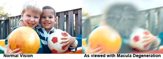
Photo Credit: National Eye Institute, National Institutes of Health
Leave a Reply