That LivingTheCRWay members want simple cost-effective ways to assess risk of Alzheimer’s Disease and dementia is quite understandable: We all want to preserve our brainpower, which is the key to quality of life and independence.
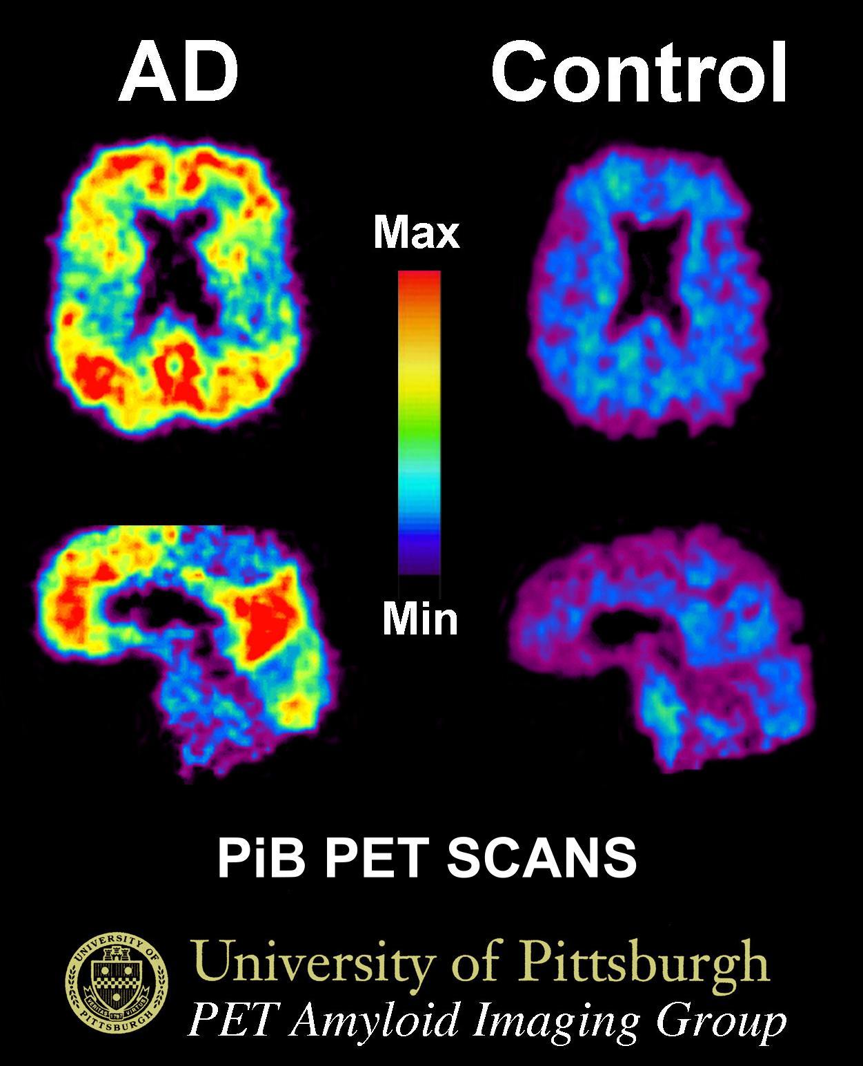 Expensive and potentially dangerous PiB-PET (Pittsburgh-compound B — Positron Emission Tomography) imaging is the gold standard for diagnosing Alzheimer’s, but it doesn’t help most LivingTheCRWay members. We do not want to be exposed to the radiation it emits. Besides, it is expensive and unavailable in many areas. That makes it impractical, especially to include as an annual test in the CR Way DNA HACR Study. Right now, the eyes seem to provide a more practical, inexpensive, and safe alternative for detecting Alzheimer’s disease years before its symptoms surface. Your eyes may also be a sensitive rate-of-aging gauge. Many research groups are studying the retina. How heartening to find that retinal vascular photography, a technique that’s available in eye centers across the country and something that many of you are already doing, may have predictive value! Consider this study:
Retinal vascular biomarkers for early detection and monitoring of Alzheimers disease
Frost S, Kanagasingam Y, Sohrabi
H, Vignarajan J, Bourgeat P, Salvado O, Villemagne V, Rowe CC, Macaulay SL,
Szoeke C, Ellis KA, Ames D, Masters CL, Rainey-Smith S, Martins RN, AIBL
Research Group
Translational Psychiatry (2013) 3,
e233; Published online
26 February 2013. doi:10.1038/tp.2012.150. e-resource
The earliest detectable change in Alzheimer’s disease (AD) is the buildup of amyloid plaque in the brain. Early detection of AD, prior to irreversible neurological damage, is important for the efficacy of current interventions as well as for the development of new treatments. Although PiB-PET imaging and CSF amyloid are the gold standards for early AD diagnosis, there are practical limitations for population screening. AD-related pathology occurs primarily in the brain, but some of the hallmarks of the disease have also been shown to occur in other tissues, including the retina, which is more accessible for imaging. Retinal vascular changes and degeneration have previously been reported in AD using optical coherence tomography and laser Doppler techniques. This report presents results from analysis of retinal photographs from AD and healthy control participants from the Australian Imaging, Biomarkers and Lifestyle (AIBL) Flagship Study of Ageing. This is the first study to investigate retinal blood vessel changes with respect to amyloid plaque burden in the brain.
We demonstrate relationships between retinal vascular parameters, neocortical brain amyloid plaque burden and AD. A number of RVPs [retinal vascular perameters] were found to be different in AD. Two of these RVPs, venular branching asymmetry factor and arteriolar length-to-diameter ratio, were also higher in healthy individuals with high plaque burden (P=0.01 and P=0.02 respectively, after false
discovery rate adjustment).
Retinal photographic analysis shows potential as an adjunct for early detection of AD or monitoring of AD-progression or response to treatments.
Read more: Keep your Glucose Low to Keep your Eyes in Good Repair
Leave a Reply